
[Everything on this website is protected by US copyright: © 2011 Novobiotronics Inc. Nothing on this website may be used without express written permission from Novobiotronics Inc.]
![]()
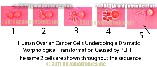
1. Two Human Ovarian Cancer Cells shown in photo( #1 above). Note the round shape of the cells.... this is the 'normal' shape for these cancer cells.
2. The cancer cell on the left begins to go through a series of 'contortions' of its shape ('morphological transformations'). It's very exciting to see this live under the microscope as it looks like the cell is 'panicking', perhaps trying to 'escape' our Plasma Emission Field Treatment (PEFT).
3. Now the cancer cell on the left is starting to fragment. The fragments still seem to 'hover' close to one another and they tend to form a kind of 'ring' of fragments surrounding what's left of the original cell, almost like many moons orbiting around a planetary body.
4. A slight change in contrast lighting under the microscope reveals that the cancer cell fragments now look less orderly, no longer circling a larger fragment, but now moving away from each other. During this stage, the cell fragments will move great distances from one another, many randomly dispersing across the microscope's field of view.
5. FINAL STAGE OF DESCTRUCTION: a remaining fragment of the cancer cell now 'hyperinflates' (arrow points to hyperinflated cancer cell remnant) and the cell no longer shows any sign of life or motion [note: cancer cells 'in-vitro' (grown in special plastic dishes for these types of experiments) commonly move around on the dish, but the speed of their movement is not easily perceived by the human eye because the cancer cells generally move rather slowly. Even cancer cells which are 'adherent' in nature (attach themselves to the bottom of the special plastic dish) will move around across the surface of the plastic dish they are 'adhered' to. ]
TIME LAPSE PHOTOGRAPHY (as used in the photos above) reveals the nature and extent of cancer cell movement during our experiments. TIME LAPSE PHOTOGRAPHTY also allows you to create 'TIME LAPSE VIDEOS' of this live action showing the morphological transformations, cell breakup and eventual 'hyperinflation'/death of the cancer cell. NOVOBIOTRONICS believes this final stage is also known as 'APOPTOSIS', or 'programmed cell death'. NOVOBIOTRONICS Inc. BELIEVES THAT PEFT CAUSES DRAMATIC CHANGES IN CANCER CELL MORPHOLOGY, EVENTUALLY LEADING TO FRAGMENTATION OF THE CANCER CELL AND TOTAL CELL DEATH [APOPTOSIS].
NOVOBIOTRONICS Inc. is currently planning additional laboratory experiments in which precise biochemical tests will be used to technically determine the onset of 'programmed cell death' ('APOPTOSIS').
Click Here to GO BACK to the Start of our Cancer Research Information